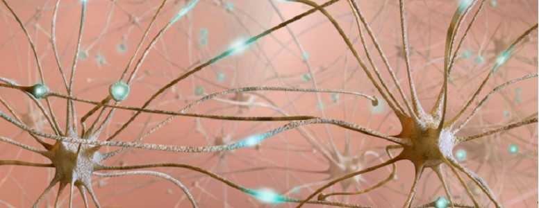
Researchers have found that a type of brain cells which surround neurons, called glial cells, can act as an appetite control switch.
The findings could have implications for therapies that help curb unbalanced eating disorders contributing to growing rates of type 2 diabetes and obesity.
Scientists long believed that glial cells served solely as supporting actors to neurons, helping them form connections with one another.
But, in the last few years, it has been suggested that glial cell activities were implicated in mediating various brain disorders.
Now, their newly discovered ability to control appetite as part of a highly regulated symphony of neurotransmitters provides clues to the development of eating disorders.
The mice study, published in the journal eLife, shows that when glial cells are activated through calcium signalling, they’re able to increase or reduce food intake by interacting with very specific neurons.
Glial cells could regulate appetite by influencing two key groups of neurons in feeding circuits of the hypothalamus, called AgRP neurons and POMC neurons.
AgRP neurons are known to stimulate appetite, while POMC neurons suppress it.
The lead author of the findings, Naiyan Che, from the Singapore Bioimaging Consortium and the McGovern Institute, tried to determine along with other MIT neuroscientists, what glial cells do to these specific neurons.
They found that the activated glial cells modulate the function of AgRPs so they hyperstimulate feeding.
However, they haven’t seen any noticeable glial-induced effects on POMC neurons, or compensatory mechanisms of glial cells when AgRP are inactivated.
This suggests that, on their ow, glial cells have strictly supporting roles, but when coupled with certain neuronal partners, that the researchers are uncovering, they can contribute to the maintenance of energy homeostasis.
A state of homeostasis is the balance between energy input, as food intake, and energy expenditure. And this was the basis for the second part of the study, which tried to see whether these interactions resulted in actual weight fluctuations in the animals.
The results show that, in the short term, mice fed ad-libitum (to satiety) who had their astrocytes, a special type of glial cells, activated, did not gain extra weight. At the same time, without any activity in the astrocytes, the mice ate less than normal.
This suggests that glial cells may cooperate with a whole bunch of other neurons, involved in energy expenditure, to compensate for increased food intake.
Appetite-related disorders may be tightly regulated in the brain, as these findings suggest, but it is important to remember that they can be largely avoided by adopting consistent healthy eating and lifestyle habits.
What's new on the forum? ⭐️
Get our free newsletters
Stay up to date with the latest news, research and breakthroughs.


