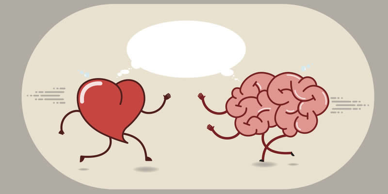

Severely stressful situations that impact the brain could lead to a broken heart, researchers have said.
A team from America has found a link between increased brain activity, caused by experiencing a stressful event, and a rare but sometimes fatal heart condition called Takotsubo syndrome (TTS).
Also known as ‘broken heart’ syndrome the muscles around the organ become weakened which leads to the left ventricle ballooning out at the bottom while the neck remains narrow.
It is a relatively new condition, which was first described in 1990. It is believed that severe emotional distress, such as grief, anger or fear, can trigger the changes to the heart.
Symptoms include chest pains and breathlessness, which in some cases can lead to fatal heart attacks. Interestingly it is more common in women than men, with only 10 per cent of cases occurring in males.
Lead researcher Dr Ahmed Tawakol, co-director of the Cardiovascular Imaging Research Center at Massachusetts General Hospital and Harvard Medical School (Boston, USA), said: “The study suggests that the increased stress-associated neurobiological activity in the amygdala, which is present years before TTS occurs, may play an important role in its development and may predict the timing of the syndrome. It may prime an individual for a heightened acute stress response that culminates in TTS.
“We also identified a significant relationship between stress-associated brain activity and bone marrow activity in these individuals. Together, the findings provide insights into a potential mechanism that may contribute to the ‘heart-brain connection’.”
The trial involved looking at scans carried out on 104 people between 2005 and 2019 at the Massachusetts General Hospital. Most of the people were being scanned to see if they had cancer, but they were also used to assess the movement of blood cells in their bone marrow.
A total of 41 people went on to develop TTS between six months and five years after the scan and 63 did not. The researchers said those who developed the condition had higher stress-related brain activity.
Dr Tawakol said: “Areas of the brain that have higher metabolic activity tend to be in greater use. Hence, higher activity in the stress-associated tissues of the brain suggests that the individual has a more active response to stress.
- High blood pressure is being overlooked in women for menopause and stress
- Females with stressful jobs associated with 21 per cent higher type 2 diabetes risk
“Similarly, higher activity in the bone marrow reflects greater bone marrow metabolism. The PET/CT scans produce images that reflect the distribution of glucose metabolism. The brain images thereby yield a map of brain metabolic activity: the higher the values, the greater the activity in those brain regions.”
The findings have been published in the European Heart Journal.


